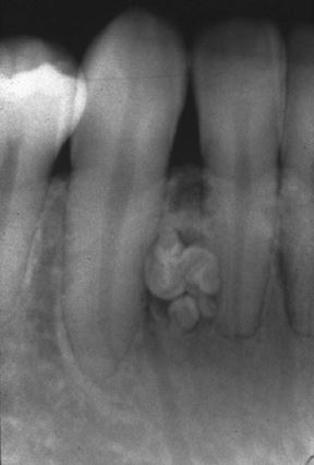
Odontoma in general refers to any tumor or growth of odontogenic origion showing complete differentiation of epithelial and mesenchymal cells. In odontomas there is formation of Enamel and Dentin due to the presence of functional Ameloblasts and Odontoblasts. As opposed to the normal Tooth, Enamel and Dentin in Odontoma are laid out in an irregular fashion which does not give the odontoma a proper shape. This happens because the odontogenic cells do not morphodifferentiate in a normal fashion. Odontoma is also called as “Composite Odontoma” as it consists of more than one type of tissue.
Odontoma or Composite Odontomas are usually smaller in size than normal teeth and are similar in presence of Enamel and Dentin.
Types of Odontomas:
- Compoud Composite Odontoma: The odontoma resembles Normal tooth in Anatomy and the way Enamel and Dentin are formed.
- Complex Composite Odontoma: The odontoma dose not resemble normal tooth in morphologic or anatomic shape, but resembles a rudimentary mass of tissue.
Etiology of Odontoma:
The basic Etiology of Odontoma is Unknown. Although not confirmed local trauma or infection during the developmental stages has been associated with Odontomas.
According to Hitchin, it has been suggested that odontomas are either inherited or are seen due to a mutant gene or interference, possible postnatal with the genetic control of tooth development.
Levy, conducted studies on Lab Rats and could successfully produce the lesion due to traumatic injury.
So the exact etiology of Odontomas is not confirmed and it is widely discussed due to no consistent location and no predilection towards maxilla or mandible.
Sex: Males > Females
Jaw: Maxilla (67%) > Mandible (33%)
Clinical Features of Odontoma:
It is mentioned in literature that Odontomas can occur in any location and at any age in any arch. But according to Budnick who analyzed a total of 298 cases of Odontomas which included 149 cases having 76 Complex and 73 Compound Odontomas along with 65 cases from Literature and remaining 84 cases from Emory university. According to Budnick, Odontomas are usually detected around the age group of 14.8 years with detection being around the second decade of life.
The location of the Odontoma differed based on the type – Compound Odontoma was found more in Anterior Maxilla region when compared to Complex type. Complex Odontomas were found more commonly in posterior maxilla region. Both Complex and Compound Odontomas were found more on the Right side of the jaw, the reason for which is not known.

Image Source: http://drgstoothpix.com/radiographic-interpretation/benign-neoplasms/odontogenic-benign-neoplasms/mixed/compound-odontoma/
Odontomas are usually Asymptomatic in the initial stages, but as the size grows slight symptoms can be seen depending on it location. Sensitivity and Discomfort to the tooth is felt if the odontoma is close to the root tip of any tooth. Expansion of bone is seen in cases where the Odontoma is associated with a Dentigerous Cyst growing around it.
Radiographic Features:
This is the most important aspect of identifying a Odontoma as most are asymptomatic and are found during routine Radiographic examinations. To identify the Odontoma we need to know that, they appear as an irregular mass of calcified material surrounded by a narrow radiolucent band with smooth outer periphery. The Odontomas are usually located between roots of two adjacent teeth.
Some cases the Odontoma might appear as a tooth morphologically mostly the crown portion and occasionally they resemble a single rooted tooth.
Developing Odontoma are difficult to diagnose, as they appear radiolucent without calcification.
Complex Composite Odontomas which are located in the posterior regions of the jaw appear as calcified mass overlying the crown of an unerrupted or impacted tooth.
Histologic Features:
Histologically we can see Enamel or enamel matrix, dentin, pulp tissue and cementum as well under the radiograph which are not arranged normally and are not related to each other in any sense.
Connective tissue capsule around the Odontoma is similar to the follicle surrounding the normal tooth during it eruption.
One Important feature is the presence of “Ghost Cells” in Odontomas which can also be seen in Calcifying odontogenic cysts.
Treatment of Odontoma:
Surgical Removal of the Odontoma is the only treatment advised.
No Recurrence is seen after surgical removal.
Microscopic examination of the Odontoma is preferred as the normal Odontoma morphologially and Radiographically resemble Ameloblastic Odontoma or Ameloblastic Fibro odontoma which have a high rate of recurrence.
Leave a Reply