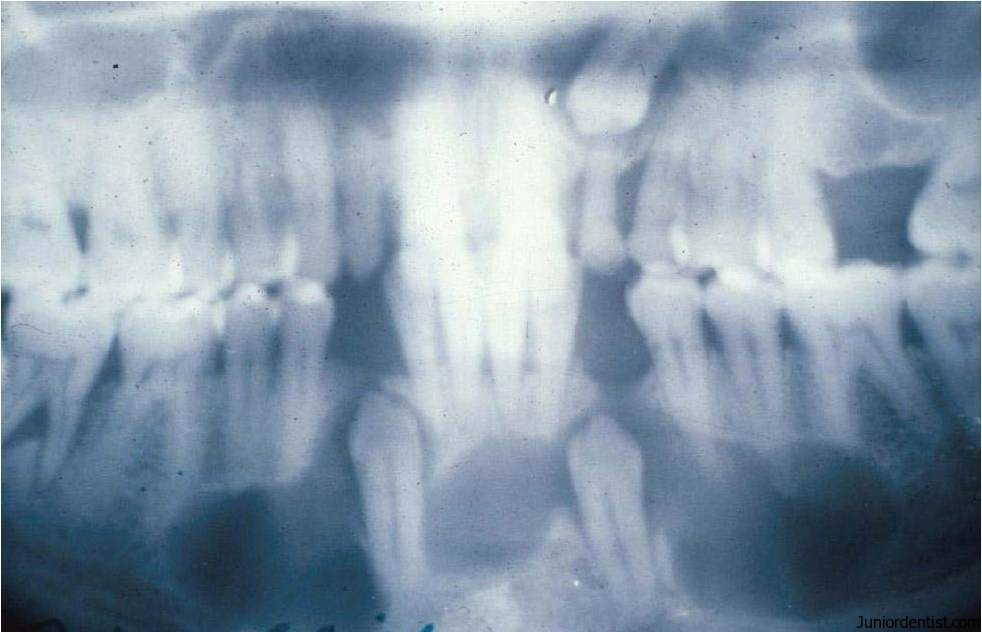
Definition: An Odontogenic cyst that surrounds the crown of impacted tooth, caused by fluid accumulation between the reduced enamel epithelium and the enamel surface, resulting in a cyst in which the crown is located within the lumen.
This is the most common developmental Odontogenic cyst.
Synonms: Follicular Cyst
Etiology:
- Develops by accumulation of fluid between the reduced enamel epithelium and the tooth crown after crown formation.
- By transformation of epithelium in the wall of the dental follicle and uniting with the follicular epithelium.
Location or Common Sites:
- Mandibular and Maxillary Third Molars and Maxillary cuspid regions
- Dentigerous cyst always is associated with the crown of a normal permanent tooth.
- Deciduous teeth are rarely involved.
Clinical Features:
It is always associated initially with the crown of an impacted, embedded or unerupted tooth
Dentigerous cyst may also be found enclosing a complex compound odontoma or imvolving a supernumerary tooth.
These are Solitary, Bilateral and Multiple cysts found usually in association with a number of syndromes like cleidocranial dysplasia and Maroteaux-Lamy Syndrome.
If Dentigerous Cyst turns into an Aggressive Lesion:
- Expansion of bone with facial asymmetry
- Extreme Displacement of teeth
- Severe root resorption
- Pain
Dentigerous Cyst may lead to Neoplasm:
- Dentigerous cyst is likely to cause Mural Ameloblastoma, which is a type of ameloblastoma.
- Localized areas of “Bud like” proliferations of cystic epithelial cells may be seen in few area, which are known as “Mural proliferations” and they indicate the development of “Ameloblastomas” which also proove the development of ameloblastoma from lining of dentigerous cyst.
- This complication will lead to difficulty in treatment of Dentigerous cyst.
Dentigerous Cyst involving an Unerupted Mandibular Third molar results in:
- Hollowing out of Entire Ramus upto the coronoid process
- Expansion of cortical plate
- Displacement of Third molar which sometimes gets compressed against the inferior border of the mandible.
Dentigerous Cyst involving an Unerupted Maxillary Cuspid:
- Expansion of the Anterior maxilla which resembles Acute Sinusitis or Cellulitis
- No pain unless Secondarily infected
Age: Seen mostly in the second and third decades of life.
Sex: Male : Female – 3:2
Radiographic Features:
- Radiolucent area is associated with un-erupted tooth crown.
- Radiolucency Symmetrically surrounds the tooth crown
- Radiolucent space will be more than 5mm
There are 3 Radiologic variants:
Central Variety: Crown is enveloped symmetrically, this pushes the crown towards the lower border of the Mandible
Lateral Type: Dilatation of the follicle on one aspect of the crown
Circumferential Type: The follicle expands in a manner which appears to envelope the Entire tooth. Sometimes the radiolucent area is surrounded by a thin sclerotic line representing bony reaction.
Histologic Features:
- Thin connective tissue wall with a layer of stratified squamous epithelium lining the lumen
-
Thin layer of epithelium, 2 – 3 layers thick with no rete ridge formation, unless infected
-
Presence of odontogenic epithelium in islands in the connective tissue wall – which may give rise to the development of AMELOBLASTOMA.
-
RUSHTON’S BODIES – peculiar, linear, often curved, hyaline bodies with variable staining of uncertain origin and unknown significance, but probably of hematogenous origin, found within the lining epithelium – especially in areas of inflammation.
-
The cystic lumen contains thin watery yellow fluid, occasionally blood stained.
-
Mandibular ones may show mucous secreting cells, sebaceous cells and lymphoid follicles sometimes.
- Rete Pegs are absent
- Connective tissue is thick and composed of fibrous tissue
Complications of Dentigerous Cyst
- Development of AMELOBLASTOMAS in the lining epithelium and in the islands of odontogenic epithelium.
- Development of MUCO EPIDERMOID CARCINOMA especially in the mandibular ones.
- EPIDERMOID CARCINOMA – due to keratin metaplasia in the epithelial component.
Treatment Of Dentigerous Cyst:
Treatment is determined by the size of the lesion
Smaller lesions: should be removed entirely- Enucleation
Larger lesions: With Bone loss and the cyst thinning the bone drastically- Marsupilisation and Surgical drainage is done.
Recurrence is rare unless there is neoplastic transformation.


its an amazing article but i want this article in my laptop in pdf plz give this with download option…..thanks for sharing this type of topics
Hello doctor Varun,
could you please send the APA citation of this article?
best regards.